Types of Breast Exams for Breast Cancer Detection
Breast cancer is the second leading cause of cancer deaths for American women, behind only skin cancer. That’s why detecting breast cancer early, while it’s most treatable, is critical.
The good news is that doctors can use many different types of breast exams to spot and assess any areas of concern. If your doctor believes you should undergo a particular test, they’ll provide details about it. But it’s a good idea to have a general understanding of the different breast examination types.
Types of Breast Examinations
What are the different types of breast exams? The most common assessment processes and tools include the following.
Breast self-exam
A breast self-exam is a check you should do regularly at home. You stand in front of a mirror and look for changes in breast size or shape, lumps, irregular thickening of tissue, and fluid leaking from your nipples.
You should also feel for lumps or abnormalities while lying down. Breast self-exams are essential and can help you spot potential issues. However, they aren’t a substitute for breast cancer screenings from a doctor or other healthcare professional. Remember that if you see or feel something unusual, it isn’t necessarily cancer. Lumps and bumps are often benign (not cancer). But, of course, you should contact your doctor about any unusual findings.
Clinical breast exam
Healthcare professionals perform clinical breast exams. These assessments involve examining both breasts for signs of cancer. The provider also checks your collarbone area and underarms. If they detect any areas of concern, they recommend imaging tests or other procedures to get more information.
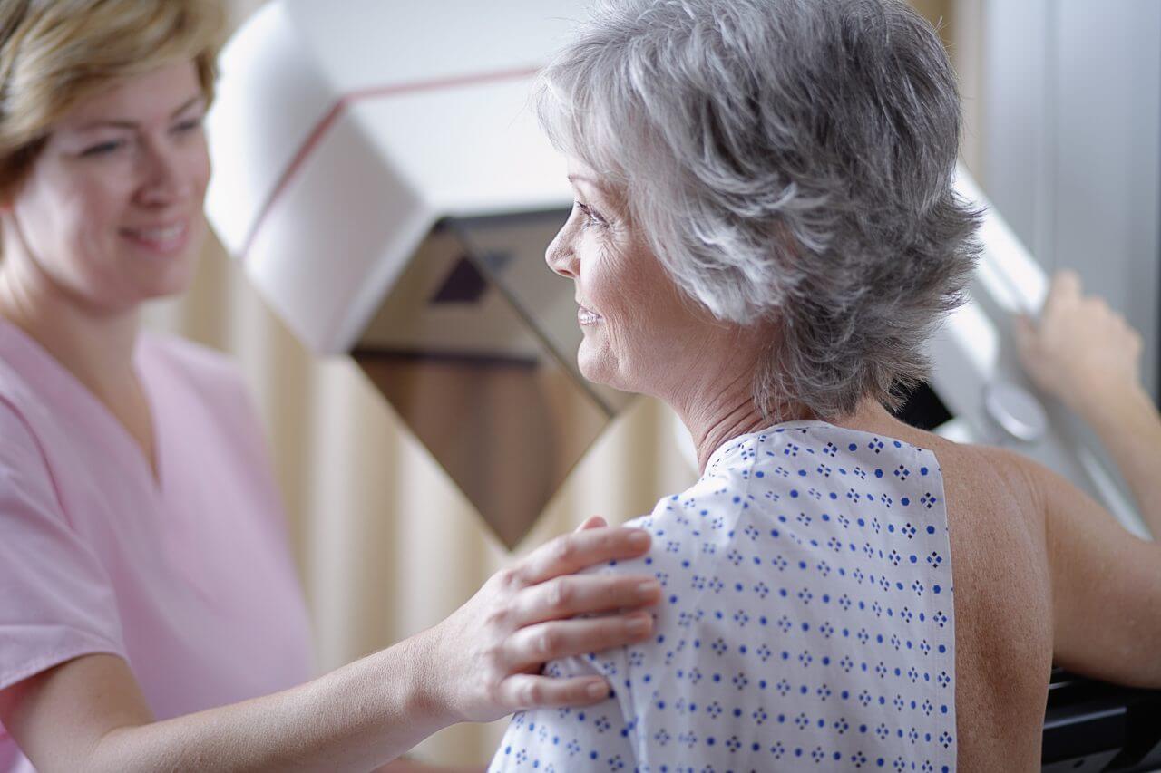
Mammogram
A mammogram is an imaging process that uses low-dose X-Rays to capture images of your breast tissue. Getting a mammogram annually or as directed by your doctor can reduce your risk of dying from breast cancer.
In a mammogram, a technician helps you position your breast between two plastic plates, which flatten it to help the machine get a clear image. The process typically involves taking two or more images of each breast, which are then reviewed by a doctor called a radiologist.
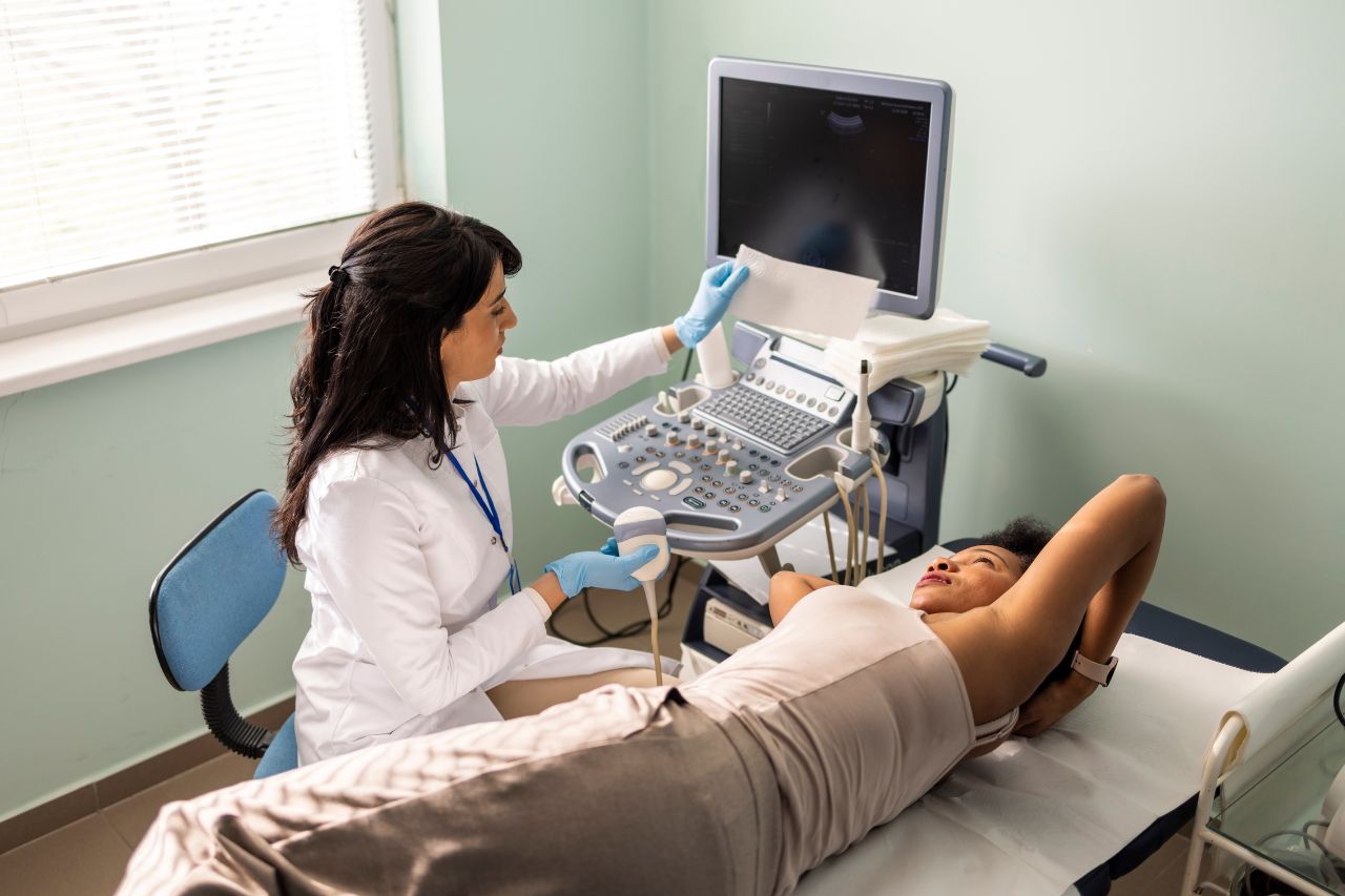
Breast ultrasound
This procedure creates images of breast tissue using sound waves. The technician places a probe against your skin, and it emits high-frequency sound waves into the tissue. The result is an image displayed on the ultrasound machine’s screen and recorded for review.
A breast ultrasound is often performed to evaluate abnormalities found in a clinical breast exam or mammogram. A radiologist interprets the results.
Breast tomosynthesis
Breast tomosynthesis is also called 3-D mammography. It’s a newer technology that takes images of the breast from different angles to create a three-dimensional picture of the tissue.
This procedure can be particularly beneficial for women who have dense breasts. A radiologist evaluates the image produced.
Contrast-enhanced digital mammography (CEDM)
This relatively new procedure is similar to a standard mammogram, but a dye called a contrast agent is injected into the breast tissue. Cancers absorb this material more readily than healthy tissue does, making it easier to identify any areas requiring additional investigation. A radiologist reviews the images generated.
Doctors sometimes recommend this procedure for women with above-average breast cancer risk or those with dense breasts.
Breast MRI
A breast MRI (magnetic resonance imaging) scan uses fluid injected into a breast, radio waves, and powerful magnets to capture pictures. A computer combines the various images into a detailed picture of the breast tissue for review by a radiologist.
This test may be appropriate for women with high cancer risk, but it isn’t typically done for those with an average risk.
Breast needle biopsy
A breast needle biopsy involves removing a small amount of breast tissue for further examination. In most instances, the area tested is not cancer, and a review of the tissue by a lab confirms that. This quick procedure causes no significant scarring and requires no recovery time.
Cyst or fine needle aspiration
This procedure can be used to remove fluid from a cyst to reduce pain or obtain cells a lab can use to confirm a lump is a cyst and not cancerous. The provider numbs the area with a local anesthetic and inserts a needle into the mass, extracting fluid and cells. This minor procedure requires no special preparation or recovery time.
Using Exams to Protect Breast Health
Breast cancer is a potentially life-threatening condition. However, combining breast self-exams, clinical exams, and imaging procedures can help ensure any problems are detected early and treated effectively.
Learn More About the Types of Breast Screening From Baptist Health
If you have questions about the different breast examination types, we’re happy to answer them. Contact your Baptist Health physician today.
You can also find helpful information on breast health in general on our website.