What Does a Pinched Nerve Look Like on an MRI?

If you experience a sharp, radiating pain, tingling or numbness, it could be a sign of a pinched nerve. When a nerve is compressed by surrounding tissues like bone, cartilage or muscle, it sends out distress signals that can disrupt your life.
While a thorough physical exam is the first step, your primary care provider may recommend a magnetic resonance imaging scan to get a clear picture of what’s happening. But what exactly does a pinched nerve look like on an MRI?
This article offers valuable insights into a common condition and the diagnostic process.
The Unseen Cause of Your Discomfort
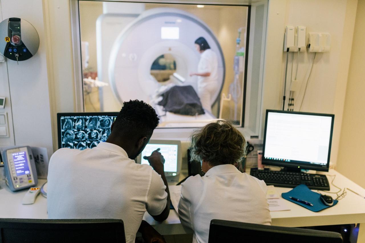
An MRI uses powerful magnets and radio waves to create detailed, cross-sectional images of your body’s internal structures. When it comes to a pinched nerve, the MRI is exceptional at visualizing the soft tissues and bones that might be causing the compression.
While the nerve itself might not always be directly visible, an MRI clearly shows the underlying issues responsible for the pain. A radiologist and your provider will look for specific issues, such as:
Herniated Discs
The soft, cushion-like discs between your vertebrae can bulge or rupture, pressing on an adjacent nerve root. An MRI can show this displacement.
Spinal Stenosis
This condition is a narrowing of the spinal canal, which can squeeze the spinal cord and nerves. The MRI will reveal a reduction in the normal space available for the nerves.
Bone Spurs
These bony growths, often caused by arthritis, can grow into the spaces where nerves exit the spine, resulting in nerve compression.
Inflammation
Swelling around a nerve, which can contribute to the pinching, may also be visible on an MRI.
Damaged nerves can also exhibit altered signal intensities, resulting in areas that look brighter or darker than the surrounding healthy tissue.
Symptoms of a Pinched Nerve
Because a pinched nerve can occur almost anywhere in the body, symptoms can vary widely. They can be intermittent or constant. The location of the pain doesn't always match the location of the pinch. For instance, a problem in your neck can cause sensations in your hand.
Common signs of a pinched nerve include:
- Sharp aching or burning pain
- Tingling or pins and needles
- Numbness
- Muscle weakness
- Hand or foot “falls asleep”
How Providers Address the Condition
After using an MRI to confirm a pinched nerve and identify its cause, your provider will develop a treatment plan. The goal is to relieve pressure on the nerve and manage your symptoms. Fortunately, many cases resolve with conservative, non-invasive approaches.
A comprehensive treatment plan is tailored to the individual and may include a combination of the following options.
Non-Surgical Treatments
- Rest and activity modification
- Physical therapy
- Splint or brace
- Ice and heat packs
- Anti-inflammatory medications
- Steroid injections
Surgical Options
If symptoms persist after several months of conservative care, surgery may be recommended to relieve the pressure. This could involve removing the part of a herniated disc or bone spur that is compressing the nerve.
Get Help With Pinched Nerves From Baptist Health
An MRI is an imaging procedure that helps healthcare providers look inside the body to identify the source of nerve pain. By revealing the precise cause of the compression, it guides an effective and targeted treatment plan, setting you on the path to recovery.
If you think or know you have a pinched nerve, consult your Baptist Health primary care provider. Call 1.877.686.1161. If you don't have a Baptist Health provider, you can find one on our provider directory.
Next Steps and Helpful Resources
Learn More About Imaging and Diagnostics
Why are MRI Machines so Loud?
How to Help Your Child Through an MRI Procedure
MRI vs MRA: What’s the Difference?


