What is Atrial Septal Defect (ASD)?
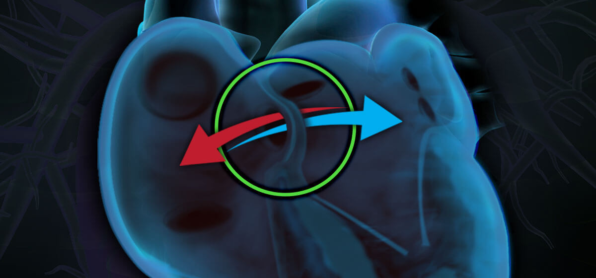
Atrial septal defect (ASD) is a hole in the septum, or muscular tissue, between the left and right atria, the upper valves of the heart. The right side of a healthy heart pumps oxygen-poor blood on to the lungs; oxygen-enriched blood is pumped from the lungs through the left atrium and throughout the body. In a heart with an atrial septal defect, the hole allows the oxygen-poor blood to mix with the oxygen-enriched blood, preventing the heart from performing its job properly.
In the case of a large ASD, oxygenated blood is pumped into the lung arteries, making the heart and lungs work harder; potentially damaging the lung arteries.
If the hole is small, the patient may not be aware of symptoms or problems. In fact, many healthy adults have a small opening in the wall between the atria.
ASDs are further categorized by location of the defect within the atrial septum. Terms used to describe defect location include secundum, primum and sinus venosus atrial septal defects. Small defects had a maximal diameter > 3 mm to < 6 mm, moderate defects measured ≥ 6 mm to < 12 mm and large defects were ≥ 12 mm.
What Causes an Atrial Septal Defect (ASD)?
As the heart forms during the first eight weeks of pregnancy, it starts as a hollow tube and develops, creating a wall between the two sides of the heart. An ASD occurs when this partition remains incomplete, leaving a hole in the heart wall.
Like most congenital heart defects, ASDs are caused by a combination of factors including family history. A mother’s condition during pregnancy, such as alcohol/drug abuse, as well as diabetes, lupus and rubella can also be contributing factors.
What are the Symptoms of ASD?
Because symptoms may not appear, patients often don’t know they have the defect. However, ASDs can show up as a murmur or irregular heartbeat, shortness of breath or fainting.
Other symptoms include:
- Fatigue after mild exercise or other activity
- Swelling in the feet, legs or abdomen
- Heart palpitations
Types of ASDs
The four most common types of ASD include:
Ostium secundum atrial septal defect – this is the most common type and affects nearly 70% of children with ASD. It occurs when part of the atrial septum doesn’t close completely.
Ostium primum atrial septal defect – is a split in the mitral valve, which controls blood flow between the left atrium and left ventricle. Further, this might occur with other congenital heart problems.
Sinus venosus atrial septal defect – this type is rarer and the defect at the connection of the superior vena cava and right atrium, where deoxygenated blood typically enters the heart. This can cause flow of already-oxygenated blood through the right atrium and back through the lungs. This is is often associated with other congenital heart problems.
Coronary sinus atrial septal defect – the rarest ASD defect, affecting the coronary sinus, where the heart’s veins feed into the right atrium.
Risks of Atrial Septal Defect (ASD)
Here are some common risks associated with ASD:
- Enlargement of the right side of the heart – can lead to heart failure.
- Abnormal heart rhythms – including atrial fibrillation or atrial flutter, affect 50- 60% of all patients over 40 with an ASD.
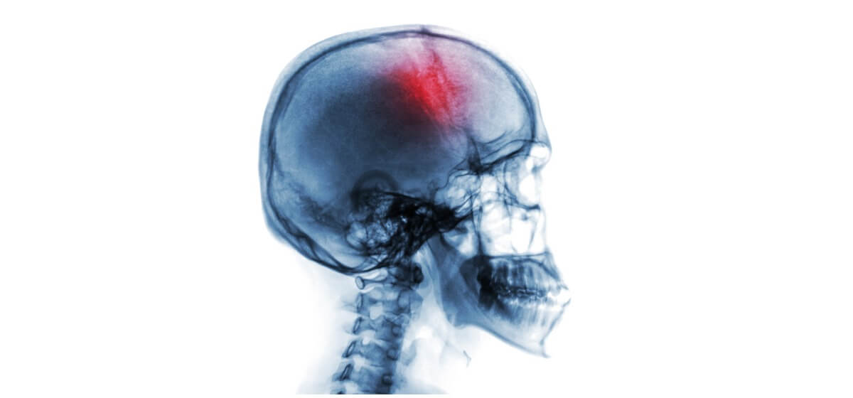
- Stroke – small clots formed on the right side of the heart are normally filtered by the lungs; when bypassing the lungs through the ASD, clots can cause stroke.
- Pulmonary hypertension – high blood pressure in the pulmonary arteries supplying blood to the lungs.
- Leaking tricuspid and mitral valves – caused by an enlarged heart.
Diagnosis of ASD
During a checkup, the detection of a heart murmur may be the first indication by your child’s doctor of a suspected heart defect or lead to an atrial septal defect diagnosis. ASDs can be diagnosed with electrocardiogram as well as X-ray and electronic imaging.
Treatment of ASD
About half of ASDs in children close on their own over time. If not, a closure device is often inserted into the hole via catheterization. Surgery is typically called for only in the rare cases of ostium primum or sinus venosus ASDs. Other options can include medical monitoring or medications. For those requiring surgery, our doctors will evaluate your condition for surgical options:
Cardiac catheterization. By inserting a catheter, or thin, flexible into a blood vessel, doctors can guide it to the heart, from the groin, and place a mesh patch or plug to close the hole. Then, the heart tissue grows around the mesh causing it to permanently seal the hole.
Open-heart surgery. Some large secundum atrial septal defects may require open-heart surgery. Through an incision, surgeons use patches to close the hole. At times, this procedure can be done using small incisions (minimally invasive surgery) and with a robot for some types.
Learn More About Atrial Septal Defect Treatment from Baptist Health
If you believe you or a loved one has atrial septal defect or would like to learn more about treatment, contact your nearest Baptist Health location today.