What Does Cancer Look Like in an MRI?
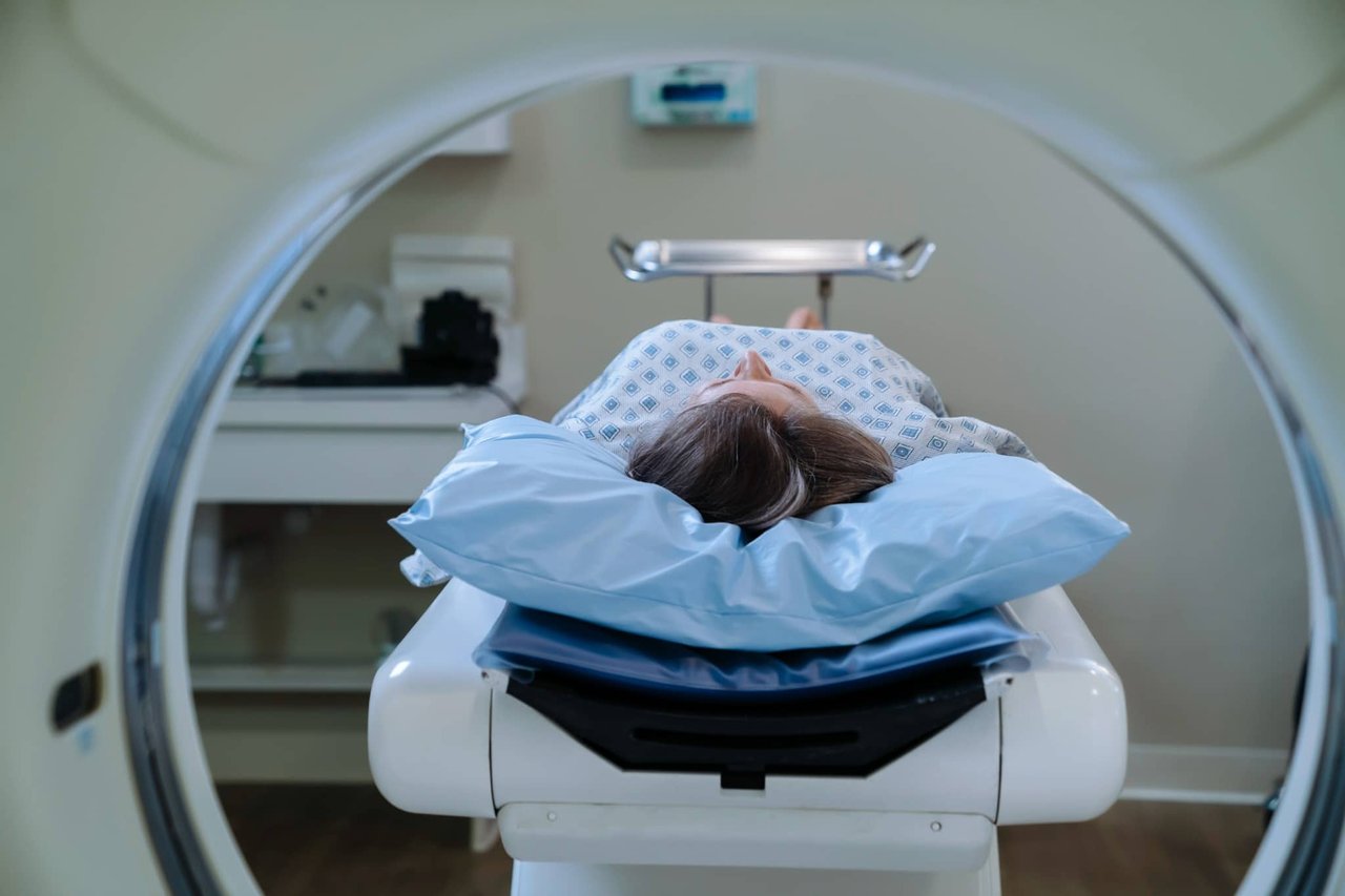
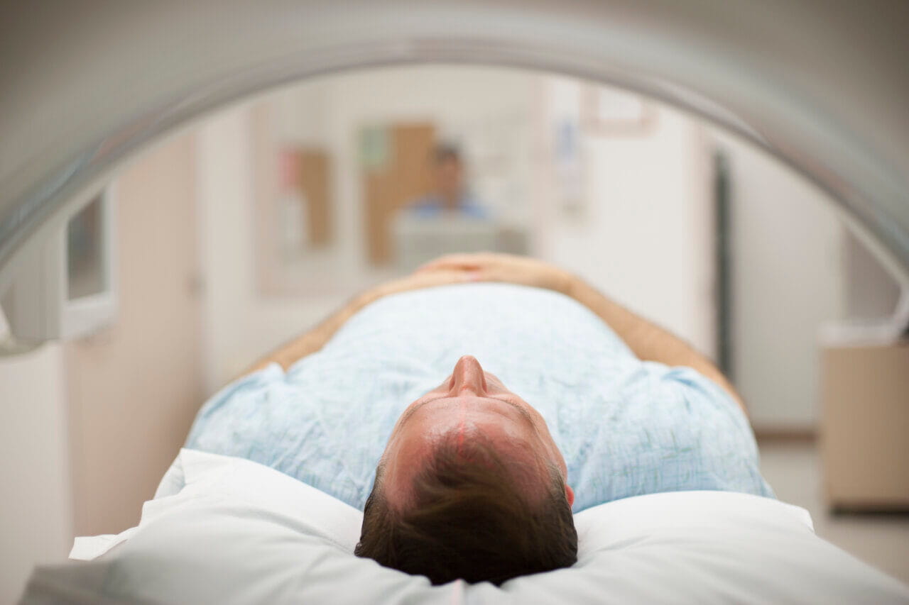
Magnetic resonance imaging, commonly known as an MRI, is a powerful tool used by healthcare professionals to visualize the inside of the body. Unlike X-rays or CT scans, this technology does not use radiation. Instead, it relies on strong magnets and radio waves to create detailed pictures of your organs and tissues. For many patients, the experience of getting an MRI can be a bit mysterious. You lie inside a large machine, hear loud noises and then wait for results.
If you’re undergoing this test to screen for or diagnose a potential health issue, you might wonder what exactly appears on those images. While the technology is complex, the goal is simple: to provide your healthcare provider with a clear view of the targeted internal structures or areas.
It’s important to remember that interpreting these images requires specialized training. A radiologist or primary care provider will review the results and explain them to you.
How the Technology Works
Knowing a little about how MRI machines create images is a first step toward understanding the results. (Another benefit is that it can help you stay calm during your procedure.) The scanner is essentially a large tube containing a very powerful magnet. When you lie inside, the magnetic field causes the hydrogen atoms in your body to align in a specific direction. The machine then sends bursts of radio waves that temporarily knock these atoms out of alignment.
When the radio waves stop, the atoms return to their original position and emit signals. A computer captures these signals and converts them into cross-sectional pictures. Because different tissues in the body—such as fat, water and muscle—respond differently to these waves, the computer can distinguish between them. This process creates a black-and-white image where different types of tissue appear as various shades of gray.
The machine creates these images in "slices," allowing providers to view the body from many angles. This level of detail makes MRI particularly good at imaging soft tissues, which can sometimes be difficult to visualize with other imaging methods.
Visualizing Abnormalities on the Scan
When a provider looks at an MRI scan, they are examining the shades of gray, white and black to identify irregularities. Healthy tissue usually has a consistent appearance, while abnormal tissue often looks different.
In many cases, cancerous tissue appears as a white or very light mass against the darker background of healthy tissue. This differs from other imaging methods, such as ultrasound, where a tumor might appear dark. The brightness or intensity of the spot on an MRI can help the provider determine the nature of the mass.
You might need to receive a contrast dye before the scan. This substance, when injected into a vein, helps certain tissues appear more clearly on images. Cancerous tumors often have an increased blood supply compared to normal tissue. As a result, they may absorb more of the contrast dye, causing them to "light up" or appear significantly brighter on the scan.
Beyond color and brightness, providers also consider the shape of the mass. Malignant or cancerous tumors often have irregular borders or shapes. They might not blend smoothly with the surrounding area. Benign or non-cancerous tumors often have smoother edges.
What Healthcare Providers Look For
It’s critical to understand that finding a "spot" or a light area on an MRI does not automatically mean cancer. Many benign conditions can also look different from normal tissue. That’s why a skilled provider must interpret the images. They look for specific characteristics to distinguish between malignant and benign growths.
Providers also use these images to determine if a cancer has spread. If a primary tumor is found, the MRI can help reveal if there are signs of cancer in nearby lymph nodes or other organs.
Types of Cancer Detected
MRI scans can help providers detect abnormalities across many parts of the body. Because an MRI excels at imaging soft tissues, it’s frequently used to examine the brain, spinal cord and nerves. It’s also highly effective for identifying tumors in the pelvic region.
This type of imaging can be used to detect and evaluate cancers in the following areas:
- Brain and spinal cord
- Breast and cervix
- Bones and joints
- Liver and pancreas
- Prostate and bladder
- Head and neck
- Colon and rectum
- Uterus and ovaries
- Soft tissue sarcomas
Your Provider Sees the “Big Picture”
While MRI images are incredibly detailed, they are just one piece of the puzzle. A provider considers your medical history, symptoms and other test results alongside the imaging. For instance, symptoms like unexplained fatigue, bleeding, weight loss or bodily function changes might prompt the need for a scan.
Ultimately, only a qualified healthcare provider can give a diagnosis. If an abnormality is found, further steps, like a biopsy, may be needed to confirm exactly what it is. The MRI helps guide that process, ensuring that any suspicious areas are investigated thoroughly.
Learn More at Baptist Health
For more information, please call the Baptist Health Oncology department at 1.855.645.1747. Visit our online provider directory to find a Baptist Health Oncologist near you. To prepare for your next appointment, take a Health Risk Assessment and download a patient packet.